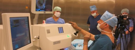
When the lens of the eye becomes cloudy, this leads to a significant visual impairment, potentially culminating in complete loss of sight. In these cases, cataract is diagnosed. Surgical procedure is the only effective way to restore the clarity of the visual path and recover visual function. There is currently no approved therapeutic treatment for cataract.
- Vision is decreased by 50 percent or more.
- Unwanted optical phenomena and decreased contrast sensitivity are causing discomfort and interfere with regular activities at home or work.
- Changes in visual perception are leading to a decreased quality of life.
- The patient sees halos around light sources.
- The disease has reached an advanced stage.
- Lack of prospects for improving functional vision (as a result of eye co-morbidity such as optic nerve atrophy, retinal detachment, or degeneration).
- Acute eye inflammation.
- Decompensated systemic co-morbidities (diabetes, systemic hypertension, rheumatoid arthritis, etc.).
- History of stroke or heart attack (within 6 months before surgical procedure).

You can make an appointment by phone from 8:30 to 19:30 (daily).
By contrast to extra- or intracapsular cataract extraction techniques, ultrasonic phacoemulsification can be carried out at any stage of the disease. This means that the patient does not have to wait until the cataract becomes mature. The cataract procedure can be done if there is substantial degradation of quality of life or if the cataract interferes with professional or daily life activities.
Some eye co-morbidities should also be taken into account. For example, in the short eye the lens may be too large, blocking the intraocular fluid outflow and leading to glaucoma. This lens is best removed, followed by intraocular lens (IOL) implantation to prevent and even cure ocular hypertension. In other cases, lens cloudiness prevents complete examination of the fundus, which is essential in diagnosing and monitoring some eye diseases. In some patients with nuclear cataracts, myopia suddenly appears that is associated with the change of the optical density of the lens. Here, surgery is needed to restore refractive status by eliminating the refractive error and the difference between the two eyes.
In some relatively young patients, there is a decrease in near vision. In such situations, the lens can be replaced with an IOL for refractive purposes, restoring vision at different distances by implanting a multifocal intraocular lens.
- Outpatient treatment or short-term in-patient stay.
- Preparations: conversation with the anaesthetist, changing clothes, taking medications if necessary.
- Local anaesthesia (with drops).
- Pupil dilation (with drops).
- Puncture of the cornea with a microsurgical blade or laser, protection of eye tissues with special solution (viscoelastic substance), insertion of the tiny ultrasonic handpiece, which is the ophthalmic surgeon’s working tool.
- Ultrasound crushing of the clouded lens, turning it into an emulsion followed by aspiration of the small particles.
- Implantation of IOL into the capsular bag.
- Evacuation of protective substance (viscoelastic solution).
- Sealing of the incision.
- Antibiotic drops or injections.
- Short medical supervision in the postoperative ward.
After 2–3 hours, without complaints and in the absence of adverse events, the patient goes home. In some cases, the patient may be advised to stay at the clinic overnight and go home the next day. The method of ultrasonic phacoemulsification is well-proven and represents the “gold standard” of ophthalmic surgery. It makes no sense to delay treatment if you can restore lost clarity of vision and quality of life with minimal risks and quick recovery.


